Transcription is a process essential to all known forms of life. To understand the mechanisms involved, we track individual proteins of interest in live cells by labelling them with fluorescent tags.
The process of transcription in micrometre-sized bacterial cells is inaccessible to traditional fluorescence imaging techniques. This constraint is imposed by the optical diffraction limit, due to the high density of fluorescent molecules emitting simultaneously which blur together signals in the acquired image. Using Photoactivated Localisation Microscopy (PALM) (1), we excite only a small subset of molecules at any given time, enabling a single molecule to be found amongst the cacophony of other molecules in the cell, and by fitting a geometric profile to the individual spots we determine its location with a precision of around 40 nm. Each molecule is then tracked by recording its position over time; and by cycling through subsets of molecules we build up a detailed picture of the location and mobility of transcription machinery (2, 3). Molecular tracks obtained from PALM are then processed to obtain diffusion properties, spatial distributions, copy numbers, and clustering properties that reveal new information about this fundamentally important biological process; initial work on live-cell applications of PALM was performed on DNA polymerase (4), and then was extended to RNA polymerase.
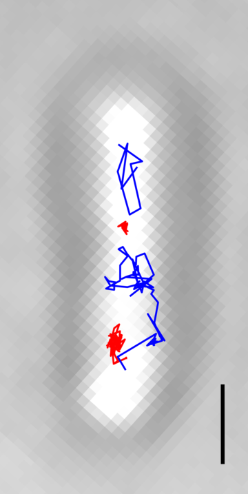
Using the PALM technique, we have investigated directly the activity of polymerases, tracking their movement during transcription. In one study we tracked RNA polymerase (RNAP) molecules in Escherichia coli cells and identified separate populations of molecules undergoing both a search process throughout the cell for transcription targets, and those engaged in transcription of the chromosome. Clustering analysis revealed that dense clusters of transcribing RNAP occur almost exclusively at the nucleoid periphery. This work provided a detailed view of the spatial organisation of transcription in living cells, and paved the way for further analysis of RNAP clustering. We have also used PALM approaches to study the mobility and spatial distribution of transcription factor, such as the lac repressor (5).
Using new purpose-built analysis tools, we are currently pushing the boundaries of our understanding of the dynamic molecular clustering of transcription machineries in live bacterial cells.
- Betzig, E. et al. (2006) ‘Imaging intracellular fluorescent proteins at nanometer resolution’, Science, 313(5793), pp. 1642–1645. doi: 10.1126/science.1127344.
- Stracy, M. et al. (2015) ‘Live-cell superresolution microscopy reveals the organization of RNA polymerase in the bacterial nucleoid’, Proceedings of the National Academy of Sciences of the United States of America, 112(32), pp. E4390–E4399. doi: 10.1073/pnas.1507592112.
- Endesfelder, U. et al. (2013) ‘Multiscale spatial organization of RNA polymerase in escherichia coli’, Biophysical Journal. Biophysical Society, 105(1), pp. 172–181. doi: 10.1016/j.bpj.2013.05.048.
- Uphoff, S. et al. (2013) ‘Single-molecule DNA repair in live bacteria.’, Proceedings of the National Academy of Sciences of the United States of America, 110(20), pp. 8063–8068. doi: 10.1073/pnas.1301804110.
- Garza de Leon, F. et al. (2017) ‘Tracking low-copy transcription factors in living bacteria: the case of the lac repressor’, Biophysical Journal. Biophysical Society, 112(7), pp. 1316–1327. doi: 10.1016/j.bpj.2017.02.028.