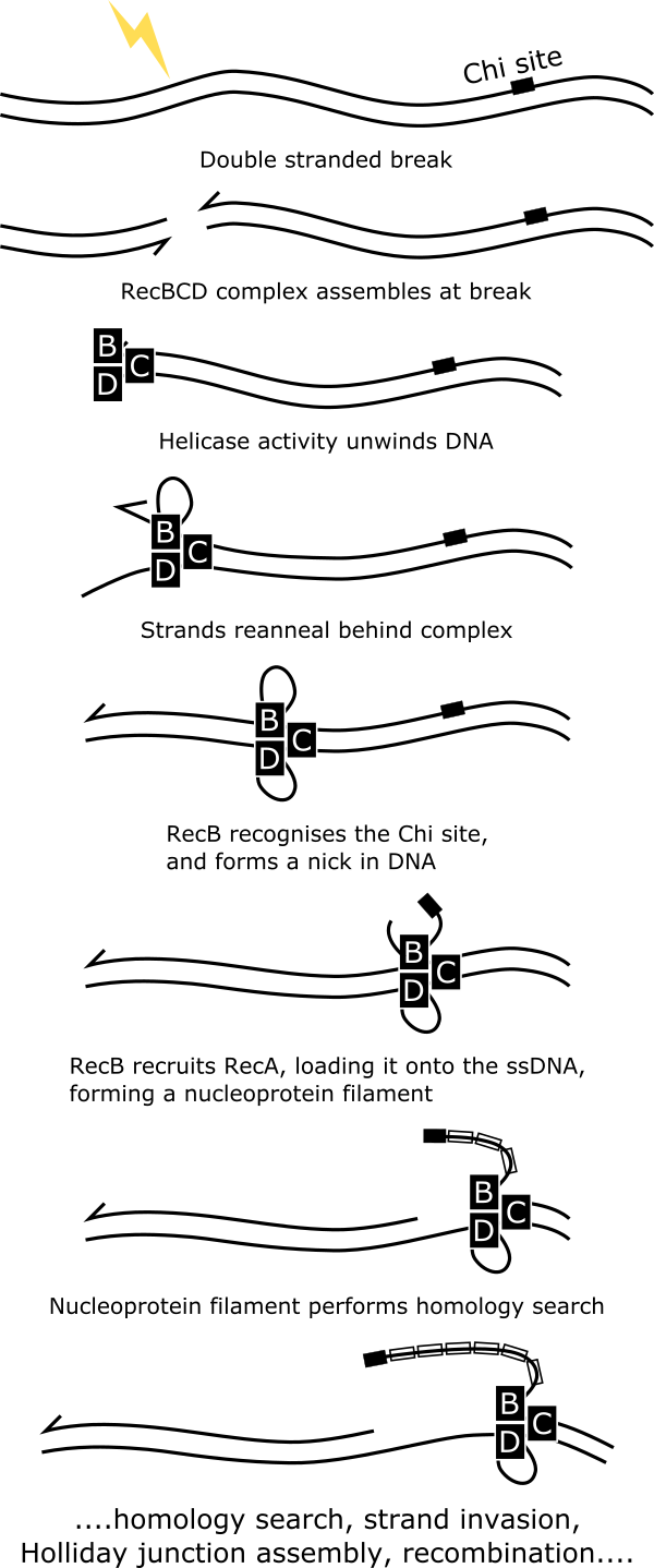
Double stranded breaks (DSBs) in which both strands of the chromosome are severed are potentially lethal to the cell. A primary mechanism used by bacteria such as Escherichia coli to repair these dangerous events is known as the RecBCD pathway. The RecBCD protein complex which initiates this process is known to identify DSBs, unwind the ends through helicase activity, and progresses along the DNA via DNA unwinding and cleavage until it identifies a sequence known as a Chi (crossover hotspot instigator) site. The RecB subunit then forms a nick in the 3’ strand, and assembles a nucleoprotein filament headed by the Chi site. Repair of the damaged region is then performed through homologous recombination.
The RecBCD complex exists in very low copy numbers in Escherichia coli (1), and important details of this vital repair pathway remain unclear. Using single molecule tracking-PALM (2, 3) we directly observe the repair process in living cells, including the complex’s search behaviour, DSB identification, and formation of the nucleoprotein filament. Using PALM we are able to quantify, and characterise each stage of the repair pathway at the single molecule level, providing insight into this critical process, the characteristics of which are often translatable into analogous repair processes in mammalian cells (4).
We have previously used the tracking-PALM technique to reveal the search process of the UvrA and UvrB proteins that perform the first steps in nucleotide excision repair functions which guard the genome against mutagenic damage from a wide range of sources (5). By modifying the bacterial genome, fluorescent labels were fused to the UvrA and UvrB proteins, which are then tracked with super-resolution precision during their repair processes. We then were able to identify an initial search process in which UvrA searches the chromosome for damage, the subsequent loading of UvrB onto the site of damage, and the dissociation of UvrA.
These experiments provide insight into important biochemical processes of DNA repair at the single molecule level in live cells, and highlight the power of super-resolution imaging techniques in this domain. The RecBCD project is a collaboration with the groups of Professor Mark Dillingham (University of Bristol), and Prof Meriem El Karoui (University of Edinburgh); the UvrA/UvrB project was a collaboration with the Sherratt group (Oxford Biochemistry).
- Lepore, A. et al. (2019) ‘Quantification of very low-abundant proteins in bacteria using the HaloTag and epi-fluorescence microscopy’, Scientific Reports, 9(1), pp. 1–9. doi: 10.1038/s41598-019-44278-0.
- Betzig, E. et al. (2006) ‘Imaging intracellular fluorescent proteins at nanometer resolution’, Science, 313(5793), pp. 1642–1645. doi: 10.1126/science.1127344.
- Stracy, M. et al. (2015) ‘Live-cell superresolution microscopy reveals the organization of RNA polymerase in the bacterial nucleoid’, Proceedings of the National Academy of Sciences of the United States of America, 112(32), pp. E4390–E4399. doi: 10.1073/pnas.1507592112.
- Lee, J. Y. et al. (2015) ‘Base triplet stepping by the Rad51/RecA family of recombinases’, Science, 349(6251), pp. 977–981. doi: 10.1126/science.aab2666.
- Stracy, M. et al. (2016) ‘Single-molecule imaging of UvrA and UvrB recruitment to DNA lesions in living Escherichia coli’, Nature Communications. Nature Publishing Group, 7, p. 12568. doi: 10.1038/ncomms12568.